Motor Neurone Disease (MND) is a rapidly progressing neurodegenerative disease that causes muscles to weaken and waste. It is a life-shortening disease for which there is limited treatment and no cure. There are currently no easy ways to detect early muscle changes in MND before major loss of function. An early sign is spontaneous muscle twitches, called fasciculations, which occur before the muscle weakens. Recent work shows that, fasciculations can show the muscles affected by MND, and the number of fasciculations per minute changes as MND progresses. This means it could be useful to measure features of fasciculation to track changes in muscle health.
Currently, detecting fasciculations is done by sticking needles into the muscles. The needles are not dangerous but can be painful and people find them unpleasant. They are also not the most sensitive way of detecting fasciculations. There is however a way of ‘seeing' fasciculations using ultrasound imaging. In previous work, we applied foreground detection-based image analysis approaches to detect fasciculations in b-mode ultrasound videos. In that we work we saw that fasciculations look different in healthy and MND affected muscles. This is promising, but further work is needed to develop ultrasound image analysis into a complete tool that can be clinically useful for earlier MND diagnosis and monitoring treatment.
This is an exciting opportunity for a fully funded fixed term research associate to work full time to advance our current ultrasound image analysis tools. This project will involve working with a collaborative team of researchers and clinicians at Manchester Metropolitan University, King's College London, and University College London, to provide a sensitive and pain-free way of monitoring muscle health in MND that will enable earlier diagnosis and support discovery of new treatments.
The Role:
The role will involve advancing biomedical image analysis and interpretation tools to improve measurement and monitoring of muscle health in people living with MND. This will involve computational analysis of previously collected ultrasound images of muscles of people living with MND and healthy controls. Our previous work is based on foreground detection using Gaussian mixture models. The work you would conduct will include using this method to analyse images and using the outputs to build, test and refine data models able to sensitively identify MND and discriminate between disease stages.
We are looking to work with someone with experience in biomedical image analysis, statistical modelling, machine learning classification and data visualization. Excellent coding skills in a suitable language is required, with knowledge of python and matlab beneficial. Experiences of working with clinical data and of industry collaboration are also desirable.
Other duties and responsibilities include:
- Maintain accurate records of research conducted and carry out analysis of the results obtained using the most appropriate method.
- Work independently and in conjunction with other investigators and collaborators.
- Prepare research findings for publication and presentation.
- Write and co-ordinate applications for research grant funding
- Assist in the supervision of postgraduate students, PhD students and Technicians.
- Participate in the activities of the research group via meetings and seminars
- Support dissemination of findings through delivery of workshops with potential stakeholders, including clinicians and industry
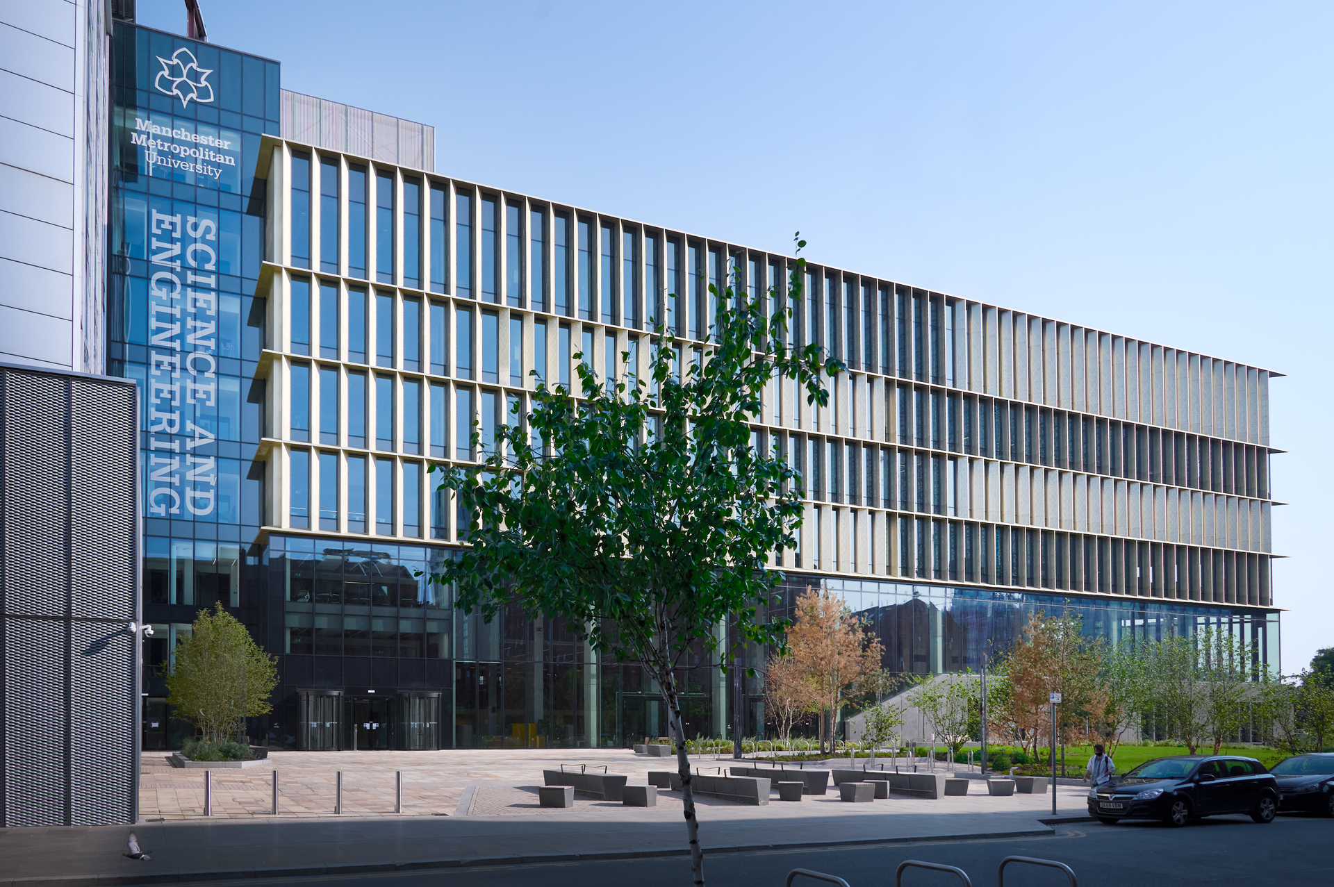
You will work within the Department of Life Sciences and Department of Computing and Mathematics, which provide state of the art facilities located in the recently built, Dalton building. The Faculty of Science and Engineering is an exciting and dynamic environment in which to conduct cutting edge research.
Qualification we require:
Hold a PhD in relevant computational or data science subjects
Application Requirements:
Experience of the following would be advantageous:
- Working with biomedical images, particularly b-mode ultrasound image sequences
- Computational image analysis, including foreground detection
- Data modelling encompassing logistic regression through to AI-based machine learning for classification.
- Different approaches to data visualisation, particularly in context of sharing with non-technical audiences (e.g., clinicians, patients, carers)
- Proficiency in statistical evaluation
- Publication in peer-reviewed journals relevant to the field
To Apply:
Please submit your CV and Cover Letter
For an informal discussion, please contact; Prof. Emma Hodson-Tole (e.tole@mmu.ac.uk).
Manchester Metropolitan University fosters an inclusive culture of belonging that promotes equity and celebrates diversity. We value a diverse workforce for the innovation and diversity of thought it brings and welcome applications from local and international communities, particularly those from Black, Asian, and Minority Ethnic backgrounds, people with disabilities, and LGBTQ+ individuals.
We support a range of flexible working arrangements, including hybrid and tailored schedules, which can be discussed with your line manager. If you require reasonable adjustments during the recruitment process or in your role, please let us know so we can provide appropriate support.
Our commitment to inclusivity includes mentoring programmes, accessibility resources and professional development opportunities to empower and support underrepresented groups.
Details
- Location:Manchester All Saints Campus
- Faculty / Function:Science & Engineering
- Salary:Grade 8 (£40,247 to £46,485)
- Closing Date:4 March 2025
- Contract Type:Fixed Term
- Contract Length:18 months
- Contracted Hours per week:37

